How Neurosurgeons Are Using the Merge Cube to Revolutionize Anatomy Education
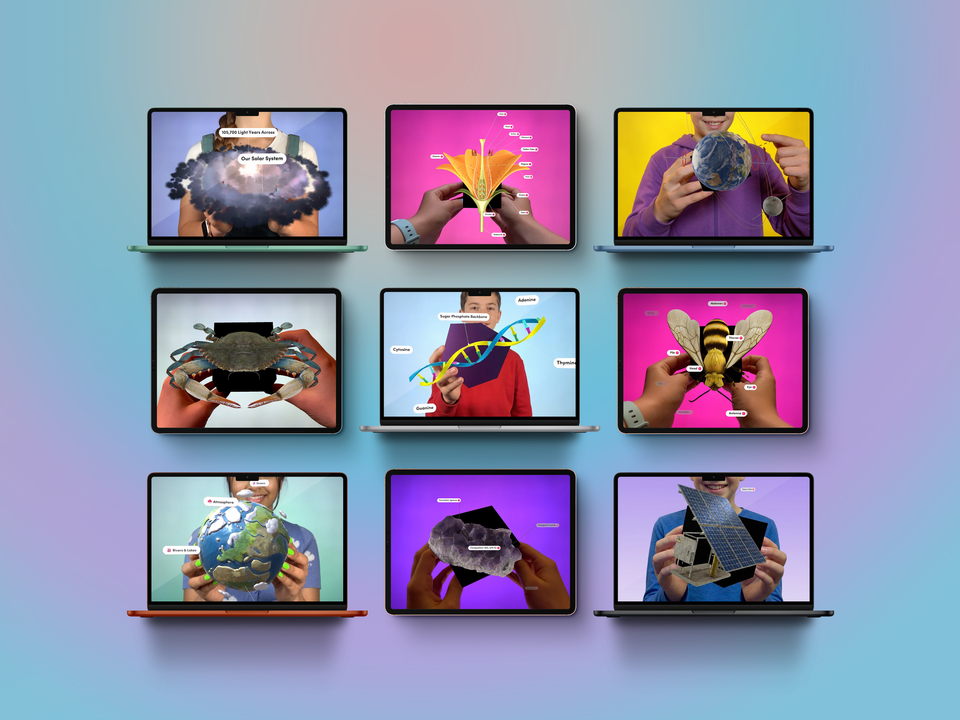
Just like young learners can explore cells, fossils, and the water cycle with Merge EDU apps, future surgeons can hold and explore anatomical models that were once confined to textbooks or inaccessible labs
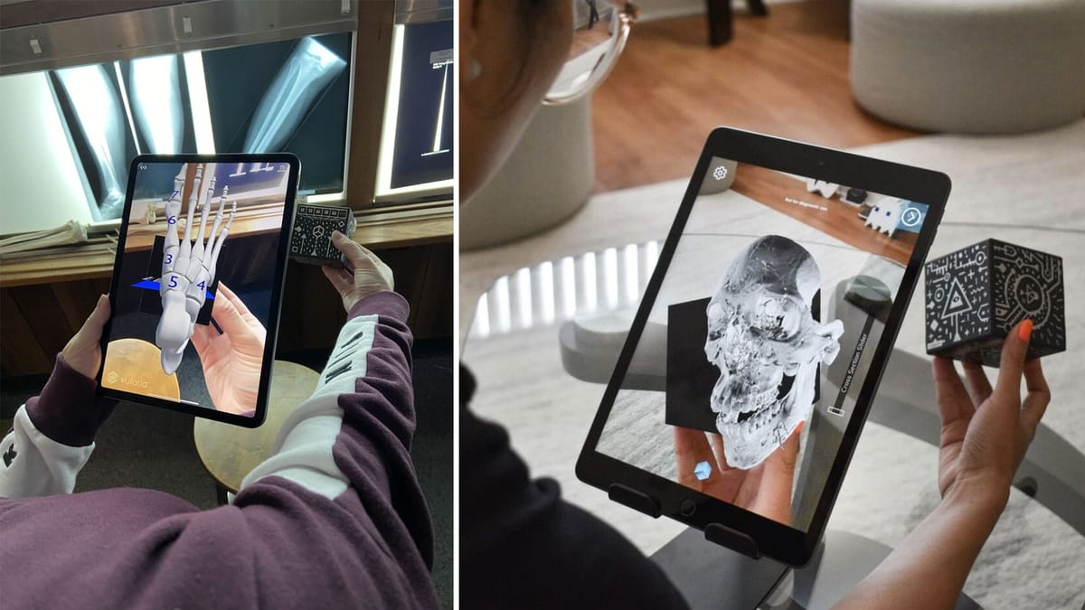
What if you could hold the human brain in your hand—and rotate it, explore it, and zoom in on intricate details—without ever stepping foot into an operating room? That’s exactly what a team of neurosurgeons from the Barrow Neurological Institute set out to do using an unexpected educational tool: the Merge Cube.
In a groundbreaking study published in the Journal of Neurosurgery (2024), researchers explored how photogrammetry and augmented reality (AR) technology can turn the Merge Cube into a portable neurosurgical lab, unlocking new ways for students and residents to engage with complex anatomy across the globe.
The Challenge of Teaching Brain Surgery
Surgical anatomy is one of the most intricate and abstract subjects in medical education. Access to cadavers is limited, and not every neurosurgery program has the resources to support high-end dissection labs. So how do we bridge that gap and bring anatomy to life in a more accessible, hands-on way?
A Cube, a Smartphone, and Photogrammetry Magic
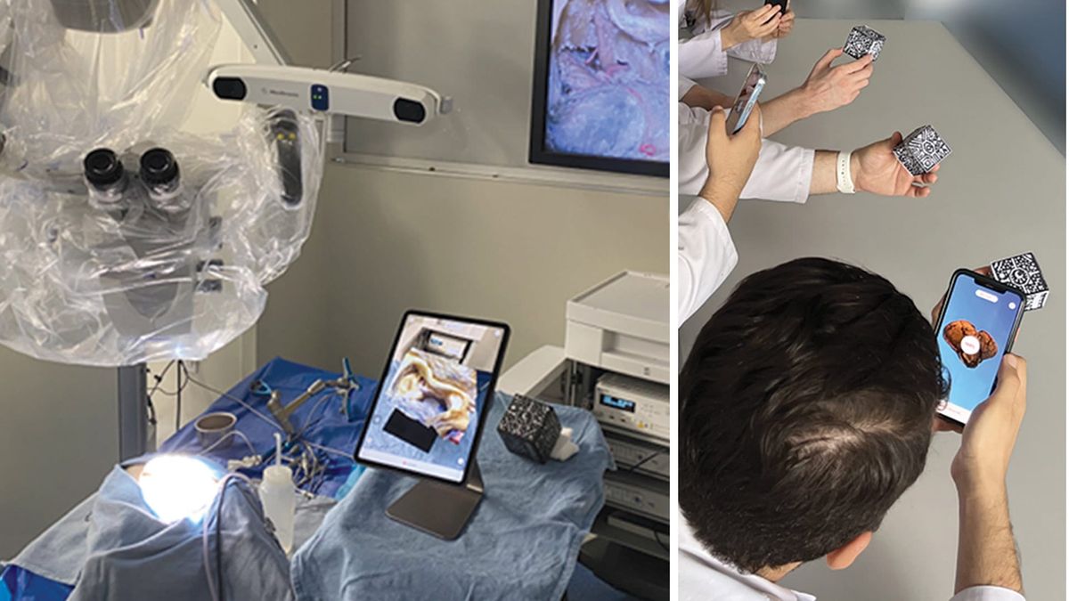
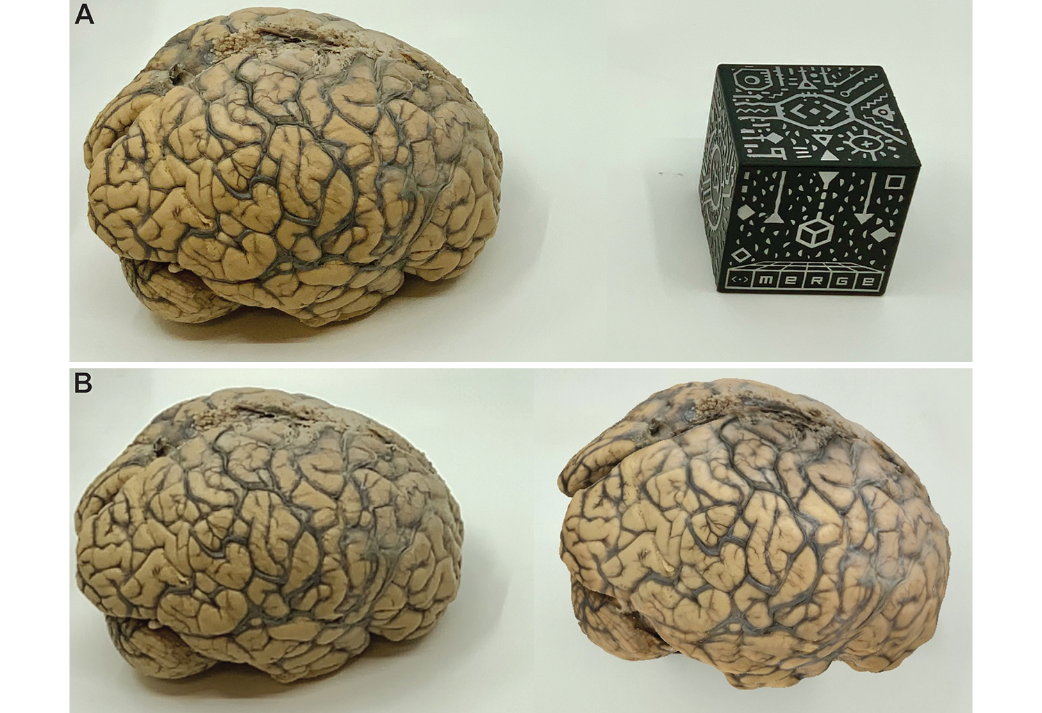
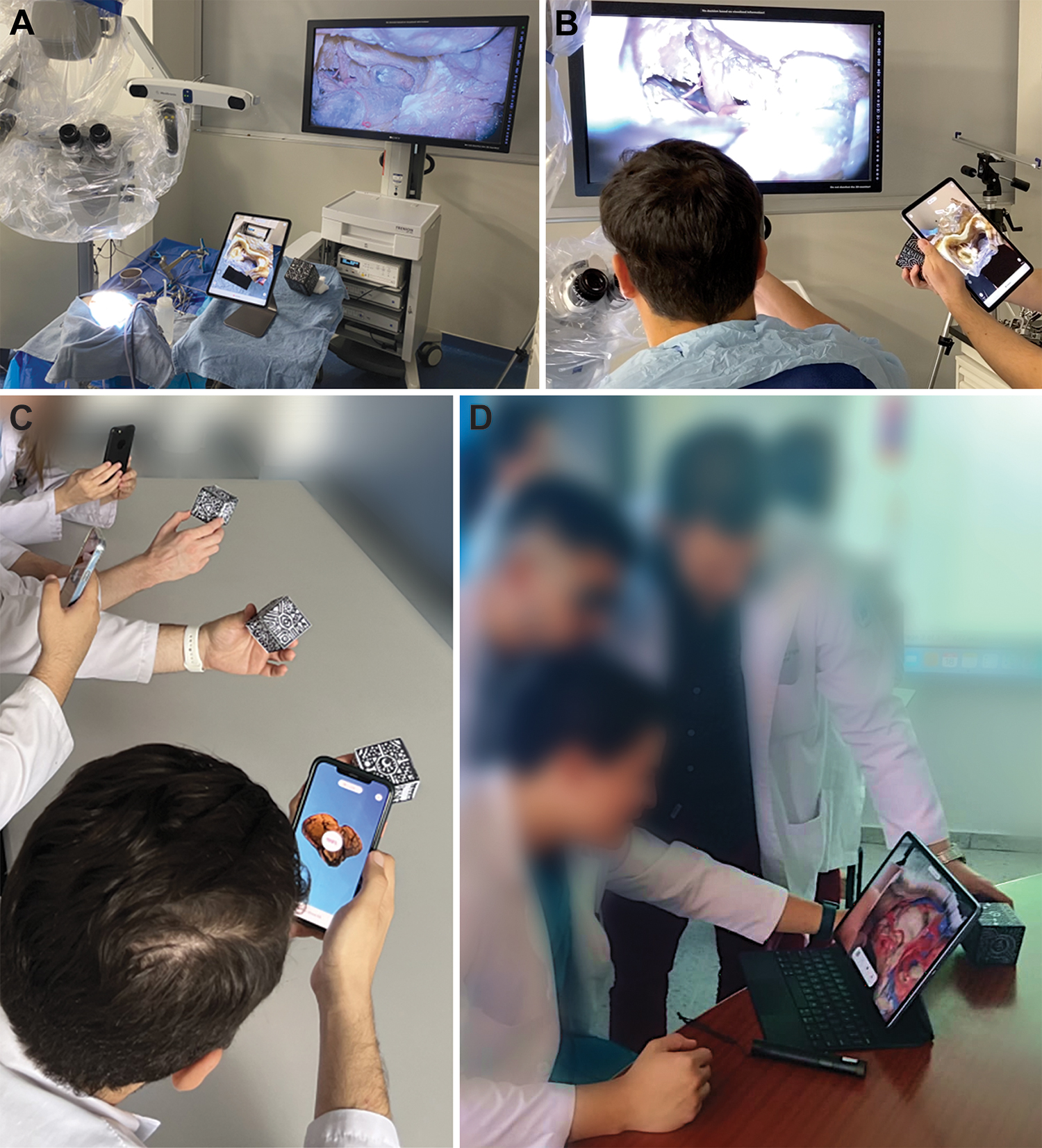
Using 3D reconstruction techniques called photogrammetry, researchers created lifelike anatomical models from cadaveric dissections. These detailed models were then embedded into the Merge Cube using scannable QR codes. With just a smartphone or tablet, trainees could explore 3D brain models from multiple angles simply by rotating the cube in their hands.
Whether it was identifying the optic carotid window or navigating the complex oculomotor corridor, users could manipulate the cube to shift surgical perspectives—just like adjusting the view through an operating microscope.
Tested Across the Globe
The AR cube system was tested in neurosurgery programs in Turkey, El Salvador, and Tanzania—locations where cadaver labs are rare or non-existent. In each setting, students and residents rated the tool highly for its educational value, ease of use, and ability to enhance their understanding of neuroanatomy.
- 94% agreed it was more engaging than traditional textbooks.
- 100% said it should be part of neurosurgical education.
- Zero reported motion sickness, visual fatigue, or discomfort.
Trainees didn’t just passively observe—they actively explored, zoomed, rotated, and learned from all angles. Instructors used the tool alongside live dissections, creating a dynamic hybrid learning experience.
Today’s Students Can Be the Creators, Too
What’s even more exciting is that students today don’t have to wait for a research fellowship or medical residency to start building with AR. With Merge Creator and the Merge Cube, students can scan real-world objects, label them with custom annotations, and present their 3D creations to the class or the world. Whether it’s a model of the heart, a handmade sculpture, or a digital replica of a community artifact, Merge empowers students to become active creators—not just consumers—of learning content. These student-built models can be used for presentations, science fairs, and even peer teaching, all while reinforcing spatial understanding and design thinking skills.
Why This Matters for Merge EDU
While this study focused on advanced neurosurgical education, the implications extend far beyond the operating room. It’s powerful validation of what we already know at Merge: hands-on AR learning works.
Just like young learners can explore cells, fossils, and the water cycle with Merge EDU apps, future surgeons can hold and explore anatomical models that were once confined to textbooks or inaccessible labs. This kind of multisensory learning—combining visual, tactile, and spatial understanding—isn’t just engaging, it’s transformative.
A Broader Shift in Medical Training
This research aligns with a broader trend we’re seeing across the medical field—augmented reality is becoming a critical tool in surgical planning, education, and practice. For example, Sira Medical is empowering surgeons with highly detailed, patient-specific 3D AR models to improve pre-surgical planning and reduce operating time. As we noted in our recent blog “Empowering Surgeons with Augmented Reality: Sira Medical’s Breakthrough”, AR isn’t just the future of learning—it’s the future of patient care, too.